The Damon Runyon Cancer Research Foundation announced that nine scientists with novel approaches to fighting cancer have been named 2019 recipients of the Damon Runyon-Rachleff Innovation Award. Initial grants of $400,000 over two years were awarded to five early career scientists whose projects have the potential to significantly impact the prevention, diagnosis and treatment of cancer. Each awardee will have the opportunity for up to two additional years of funding (four years total for $800,000). In addition, continued “Stage 2” support was granted to four awardees, who demonstrated significant progress on their proposed research during the first two years of the award. This year, the Foundation increased the award by 33% from $150,000 to $200,000 annually.
The Damon Runyon-Rachleff Innovation Award funds cancer research by exceptionally creative thinkers with “high-risk/high-reward” ideas who lack sufficient preliminary data to obtain traditional funding. The awardees are selected through a highly competitive and rigorous process by a scientific committee comprised of leading cancer researchers who are innovators themselves. Only those scientists with a clear vision and passion for cancer research are selected to receive the prestigious award. Examples of past success stories from Damon Runyon awardees include the gene editing technology CRISPR and single cell sequencing techniques that are revolutionizing not just cancer research, but biomedical research globally.
This program was established thanks to the generosity of Andy and Debbie Rachleff.
2019 Damon Runyon-Rachleff Innovators:
 Xiaochun Li, PhD, The University of Texas Southwestern Medical Center
Dr. Li is focusing on the Hedgehog (Hh) signaling pathway, which is required for proper development during the formation of an embryo. This pathway can also be activated abnormally in adult tissue and has been implicated in multiple cancers—including basal cell carcinoma and medulloblastoma—that account for about 25% of cancer deaths. His lab will apply structural and cell biological approaches to study two major components of the Hedgehog signaling pathway: Patched-I, a tumor suppressor, and Smoothened, an oncoprotein. These results will broaden the understanding of the Hh pathway in cancer progression and may help identify potential targets for therapeutic intervention.
 Joseph D. Mancias, MD, PhD, Dana-Farber Cancer Institute
Dr. Mancias is exploring the synergistic interactions between radiation therapy and targeted immunotherapy in patients with pancreatic cancer (pancreatic ductal adenocarcinoma, “PDAC”). While this treatment combination has shown dramatic benefits for patients with certain cancer types, it has been challenging to predict which patients will respond and to determine how to harness the anti-tumor immune cell effects of radiation. Identifying the PDAC radiation-induced antigens will shed light on the interaction between the immune system and radiation and provide the basis for designing effective immunotherapies for patients with pancreatic and other cancers (such as glioblastoma, prostate cancer, breast cancer, lung cancer, head and neck cancers, pediatric cancers).
 Jan P. Schuemann, PhD, Massachusetts General Hospital
Though radiation therapy is a key treatment for many solid tumors, healthy surrounding tissue can be damaged through inadvertent exposure leading to serious side effects. Dr. Schuemann’s research is exploring if “extreme dose rate (EDR) proton therapy” can be used to spare healthy tissue while treating solid tumors. With new EDR technology, proton radiation therapy is delivered extremely fast—whole treatments in milliseconds. However, the mechanism underlying the reduction in side effects is still unknown. This research will focus on brain irradiations but has the potential to be applied to nearly all cancer types. These results may demonstrate how EDR treatments can greatly improve radiation therapy outcome.
 Jason M. Sheltzer, PhD, Cold Spring Harbor Laboratory
Dr. Sheltzer studies how aneuploidy, or having too many or too few chromosomes in the cell, affects cancer development and treatment. Approximately 55% of breast cancers have an extra copy of one part (called the “q arm”) of chromosome 1. His lab is developing cutting-edge chromosome engineering technology to eliminate the extra copies of 1q from breast cancer cell lines and determine whether this prevents the cells from forming tumors. Additionally, his group will test whether aneuploidy causes ovarian cancer cells to be sensitive to any chemotherapies, with the goal of identifying a drug that specifically kills cells with extra copies of chromosome 1q without affecting normal cells. These experiments could lead to highly effective “chromosome-specific” therapies based on aneuploidy.
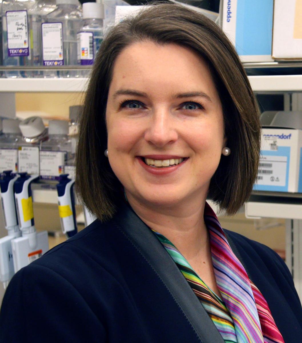 Alexandra-Chloé Villani, PhD, Massachusetts General Hospital
Immune checkpoint inhibitors unleash the immune system to attack tumors; they have revolutionized the treatment of solid cancers by changing the prognosis for many patients, improving their quality of life and offering long-lasting remission. However, these immunotherapies can also spur assaults on healthy organs called “immune-related adverse events” (irAEs), ranging from minor rashes and fevers to severe gastrointestinal complications and deadly heart inflammation. Dr. Villani is analyzing patient samples using state-of-the-art genomic technologies and integrative immunological approaches to understand why and how these irAEs occur in cancer patients. Ultimately, she aims to identify therapeutic solutions to prevent or clinically manage irAEs without reducing the lifesaving potential of immunotherapy.
2019 Stage 2 Damon Runyon-Rachleff Innovators:
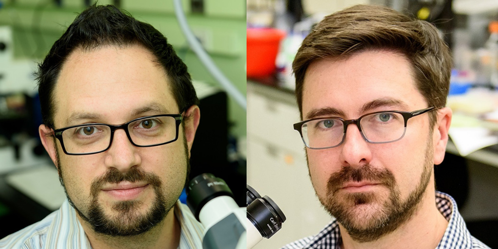 Benjamin L. Martin, PhD and David S. Matus, PhD, Stony Brook University
Metastasis, when tumor cells spread to distant organs and form secondary tumors, is one of the most deadly aspects of cancer but is not well understood. Drs. Martin and Matus are focusing on this process to understand how cancer cells break free from tumors to move through the body. Their collaborative project is founded upon an experimental observation made by Dr. Matus in the model roundworm, C. elegans, that a cell cannot simultaneously invade and divide. Using two model systems, C. elegans and the zebrafish, D. rerio, they have gained a better mechanistic understanding of how cell cycle arrest increases the invasive capacity of individual cells during metastasis, including extravasation, the ability to exit blood vessels into the surrounding tissue. They have developed a biosensor that allows the quantification of cells at different stages of the cell cycle and specialized light sheet microscopes to visualize invasive cancer cell behavior at high resolution, in real time. Insights from their work may aid in the design of therapeutics to eradicate metastatic cells that escape traditional chemotherapeutic agents which only target actively dividing cells.
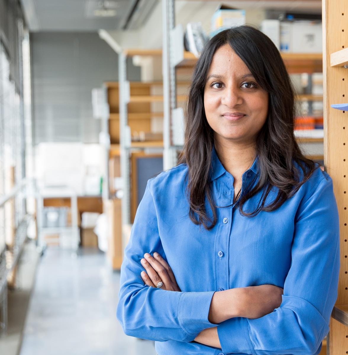 Rushika M. Perera, PhD, University of California, San Francisco
Cancer cells have a unique ability to rapidly and efficiently remodel their internal composition and metabolic pathways in order to maintain accelerated growth, metastasize and resist anti-cancer therapies. The lysosome, an organelle in the cell that degrades cellular debris, has the ability to control a cancer cell’s adaptability. Through processing and recycling different macromolecules, the lysosome serves as an important source of fuel for cancer cell growth and spare parts for remodeling the cell. Dr. Perera is focusing on pancreatic ductal adenocarcinoma (PDAC), which is highly dependent on lysosomes for growth. She has characterized cancer-specific lysosomal proteins and showed that these proteins confer PDAC cells with two key properties: the ability to rapidly repair their membranes in the face of sustained mechanical and chemical insults, in order to maximize nutrient uptake, and to alter their cell membrane composition to evade recognition by the host immune system. Collectively, these results suggest largely unexplored roles for the lysosome in PDAC and highlight novel vulnerabilities that could be exploited therapeutically.
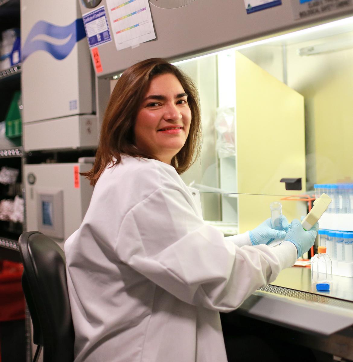 Marcela V. Maus, MD, PhD., Massachusetts General Hospital
Dr. Maus is engineering the body’s own immune T cells to fight deadly brain tumors like glioblastoma. However, in studies of patients with brain tumors, she has found that tumor cells can escape the engineered T cells. She is now redesigning T cells so that they block escape routes used by the tumors. She expects that the engineered cells will be more powerful and may become a new effective treatment for brain tumors. The engineered CAR T cells are designed to target cells displaying multiple abnormal proteins (antigens) made from cancer-causing oncogenes; theywill act as drug carriers to address the specific hurdles of antigen heterogeneity and penetrating the blood brain barrier. She is testing these “living drugs” in vitro and in mouse models with the goal of ultimately advancing these studies to human clinical trials. Furthermore, if this system works for brain tumors, it has the potential to be applied as a therapy for other forms of cancer as well.