The outermost layer of the human brain, known as the cerebral cortex, is responsible for our highest mental capacities—language, memory, emotion, decision-making, and much more. It contains an immense diversity of cells, between 14 and 16 billion neurons, organized in patterns complex enough to elude the farthest reaches of neuroscience.
With the rise of single-cell RNA-sequencing, however, it is now possible to identify distinct brain cell types and pinpoint their location in the tissue, resulting in “cell atlases” that better capture the diversity and spatial organization of a given region. These maps not only provide insight into normal brain function but also help us understand where and how problems occur in neurodegenerative diseases.
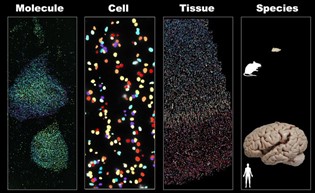 At Harvard University, this is what Damon Runyon Fellow Rongxin Fang, PhD, and his colleagues are doing with the middle and superior temporal gyrus, areas of the brain responsible for language comprehension and facial recognition, among other processes. Using single-cell sequencing tools and an advanced imaging technique called MERFISH, the team has generated a cell atlas of these regions that includes over 100 distinct cell types. For comparison, they also performed MERFISH imaging in multiple cortical areas in the mouse brain.
As we might expect, Dr. Fang and his team found major differences in both the cellular composition and organization of the human and mouse brains. For example, humans have a much higher proportion of glial cells, non-neuron cells that protect and support neurons. Human neurons also have more proximity to and interactions with glial cells, and particularly with oligodendrocytes, which wrap the axons of neurons in a lipid-rich material called a myelin sheath. This comparatively high level of support and protection is due, the researchers posit, to “the higher energy demands of firing a single neuron in the human brain” compared to that of a mouse.
The team also observed a high degree of proximity and contact between specific glia-neuron pairs within the human cortex. Interestingly, some of the ligand-receptor pairs in these cells are genetically associated with neurodegenerative diseases, indicating a possible molecular basis for neurodegeneration.
With these findings, Dr. Fang demonstrates the utility of genome-scale imaging, a technique he is also using to study gene expression in cancer. This cross-species spatial analysis of the brain, as the first of its kind, offers hope that other blurry areas of human biology will soon sharpen into single-cell resolution.
This research was published in Science.