Paul C. Klauser, PhD
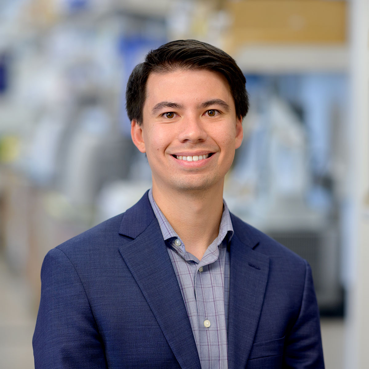
Radiopharmaceuticals, or drugs that contain radioactive forms of chemical elements, have transformed cancer diagnosis and treatment. Radioactive copper and manganese, for example, play a crucial role in PET imaging, while radioactive lutetium is used to deliver targeted radiation to cancer cells. While these radiometals have tremendous potential, however, their application is hindered by a lack of efficient “chelators,” or molecules that can securely bind radiometals in the human body. Computational protein design offers a solution by engineering protein-based chelators optimized for radiometal coordination, stability, and biocompatibility. Using advanced protein modeling, Dr. Klauser [Marilyn and Scott Urdang Quantitative Biology Fellow] will develop chelators for radiometals, improving diagnostic imaging and advancing lutetium-based radiotherapies. While this work is applied to HER2-positive gastric cancer, these strategies have broad applications across various cancer types, ultimately enhancing precision oncology and expanding radiopharmaceutical utility.
This research develops a computational strategy to design stable, compact metal-binding proteins for radiopharmaceuticals, enabling fusion with therapeutic antibodies. Using diffusion models, such as RFdiffusion, thousands of protein backbones are generated for metals like copper and manganese. Sequences are assigned via ProteinMPNN, filtered for stability and binding with AlphaFold 3. For rare lanthanides, symmetric duplication of known binding motifs is used. This approach streamlines the discovery of stable scaffolds for radiopharmaceutical applications.
Ruoyu Wang, PhD
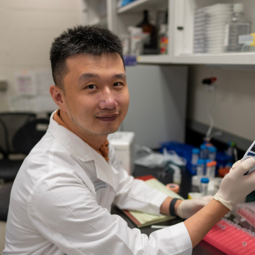
Many cancer mutations occur in regions of the human genome that do not code for proteins. These non-coding regions serve as vital regulators of gene expression; mutations in these regions contribute to various hallmarks of cancer. Elucidating these regulatory elements and their malignant variants is critical for advancing our understanding of cancer biology and fostering precision medicine. Deep learning sequence models can substantially enhance our grasp of the regulatory genome in both health and disease. To this end, Dr. Wang aims to combine generative AI models with single-molecule regulatory genomics to uncover the principles that underlie the cancer regulatory genome at unprecedented resolution and precision.
With single-molecule regulatory genomics, Dr. Wang will develop a deep generative AI model to learn the probability landscape of the single-molecule regulatory genome. By taking any DNA sequence as input, the deep generative AI model can generate diverse configurations of single-molecule chromatin states.
Simone Bruno, PhD

Triple-negative breast cancer (TNBC) is one of the most aggressive and difficult-to-treat forms of breast cancer. Dr. Bruno’s research focuses on how changes in DNA packaging, known as chromatin, affect cancer progression and treatment response. Using advanced computational models, Dr. Bruno will investigate the interplay between chromatin modifications and resistance mechanisms, with the goal of identifying strategies to mitigate therapy resistance and reduce cancer recurrence. While Dr. Bruno’s primary focus is TNBC, this research provides a generalizable framework that could be applied to other cancers where chromatin modifications play a key role, leading to better, more durable treatments for a broader range of patients.
In this project, Dr. Bruno will develop a computational framework that integrates mathematical models of chromatin modification dynamics with pharmacokinetic and pharmacodynamic models to study the role of these modifications in triple-negative breast cancer (TNBC) progression. Dr. Bruno will adapt existing chromatin modification circuit models to TNBC dynamics and, using Bayesian inference methods, will parameterize them with experimental data. By integrating pharmacokinetic models for specific drugs, this framework will enable the evaluation of diverse treatment strategies.
Sohyeon Park, PhD
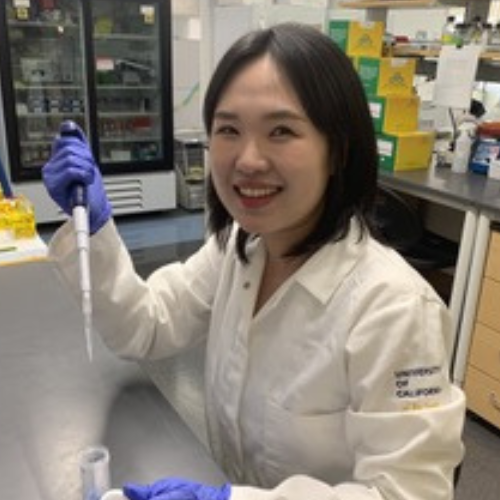
Macrophages are a major component of the body’s first line of defense, acting as sentinel cells that detect and respond to threats. One powerful trait of macrophages is their ability to modulate their response based on previous exposure to stimuli. In cancer, this adaptability can steer macrophages either to fight tumors or to protect them, depending on prior experiences. This phenomenon is often referred to as “macrophage memory.” Though macrophage memory is increasingly recognized as a factor influencing cancer progression and treatment outcomes, the mechanisms that allow macrophages to retain this memory remain unclear. Dr. Park hypothesizes that exposure of macrophages to certain stimuli leads to lasting changes in the structure of their DNA. She will combine both experimental and computational approaches to elucidate how this memory forms and how it affects the expression of immune-related genes. By uncovering the principles of macrophage memory formation, she will lay the groundwork for strategies to reprogram macrophages, potentially enhancing anti-tumor immunity and improving cancer therapy.
Dr. Park will use machine learning–aided genome modeling, leveraging bulk Hi-C data, to reconstruct 3D chromosome structures and validate them with deep learning–based image analysis. This framework will infer single-cell 3D chromosome structure in macrophages. Dr. Park will also quantify nuclear speckle–mRNA spatial relationships using microscopy and develop mathematical models of gene regulation that incorporate transcription factor activity. This integrative approach will reveal how chromosome structure shapes macrophage immune memory and functional response in cancer.
Aaron Zweig, PhD
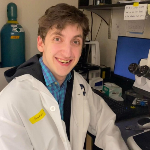
Dr. Zweig [Sijbrandij Foundation Quantitative Biology Fellow] is modeling how gene regulation changes across time and space using advanced geometry, deep learning models, and CRISPR gene-editing technology. With these tools, it is possible to learn how RNA expression evolves in a cell over time and, ideally, predict evolution in regions susceptible to cancerous mutation(s). This work has the potential to apply to many types of cancer but is especially applicable for acute myeloid leukemia (AML). This is in part because there are known “precursor” states—when cells are at higher risk of becoming cancerous—where understanding gene regulation is most impactful, and in part because a common element of AML treatment is a stem cell transplant, which may attack host cells inconsistently in different regions of the same organ.
The project models temporal gene dynamics via stochastic differential equations, parameterizing the drift vector field with linear networks or shallow neural networks to guarantee provable identifiability, trained via differentiation through the adjoint method. Spatial interactions between spot clusters are characterized with graph neural networks, symmetric functions parameterizing summary statistics, and self-attention modules on latent gene embeddings. The latent embeddings are also defined through a variational autoencoder integrating RNA with other modalities.
Ahmed Roman, PhD
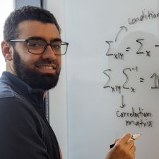
Dr. Roman [Leslie Cohen Seidman Quantitative Biology Fellow] aims to develop mathematical tools to determine which genes are associated with resistance to chemotherapy. Given genomic information from pancreatic cancer patients whose tumors are resistant or sensitive to chemotherapy, this tool will identify genes that distinguish the two populations. These genes can then be explored as potential drug targets that can sensitize chemotherapy-resistant tumors to treatment.
Dr. Roman’s research relies on the use of information theory to improve the ability of neural networks to find genes whose RNA expression distinguishes chemotherapy-sensitive from resistant patients. Another research direction is to leverage prior knowledge, accumulated over decades about gene-gene interactions in the laboratory, to inform the architecture of the neural networks or use large foundation models training on millions of cells to study cancer.
Jeremy A. Owen, PhD
Chromatin remodelers are complex protein machines responsible for packaging DNA and regulating gene expression. Their dysfunction is strongly implicated in cancer. For example, certain types of sarcoma and ovarian cancer are driven by mutations in a chromatin remodeler called BAF. Combining experiments with theoretical work, Dr. Owen’s research aims to understand how remodelers recognize their target sites in the cell’s nucleus. By expanding our understanding of chromatin remodeling, the findings of this research will provide the groundwork for more effective cancer treatments—suggesting how drugs might target chromatin remodelers—as well as enhance our understanding of how existing drugs that target remodeler-adjacent mechanisms might work.
A central aim of this project is the development of new, quantitative models to explain the behavior of chromatin remodelers seen in experiments. Dr. Owen will achieve this by successive rounds of passing between theory and experiments repeatedly—measuring, modeling, then measuring again. For comparison to experiments, model predictions will be extracted computationally (e.g., numerically solving ODEs, or by exact stochastic simulation using Gillespie’s algorithm) or analytically (e.g., by the King-Altman procedure, and variants), as appropriate.
Isabella N. Grabski, PhD
Only 3% of cancer drugs in clinical trials ultimately receive FDA approval, compared to 15-33% of drugs for other types of diseases. Recent studies have suggested that many drugs being explored for cancer treatment do not actually target their intended molecule in the cell. This has important implications for efficacy and safety and could be a key contributor to the low FDA approval rate. Dr. Grabski [Kenneth G. Langone Quantitative Biology Fellow] has created a novel experimental and computational framework to identify drug mechanisms of action at molecular resolution by leveraging CRISPR-based technologies. With this framework, she hopes to more precisely identify how a given cancer drug functions in the cell. This could serve as a powerful tool for preclinical evaluation and even potential discovery of new cancer therapeutics.
Dr. Grabski’s project aims to identify drug targets by modeling drug transcriptional response as a sum of genetic perturbation responses. She will perform this deconvolution in two steps. First, she will use a multi-condition latent factor model to produce denoised estimates of perturbation effects. Second, she will leverage sparse Bayesian regression techniques to map drug responses to these perturbation effects, in a way that can summarize complex patterns of uncertainty among related perturbations.
Carolina Trenado-Yuste, PhD
Breast cancer is the most frequent cancer in women and the second-leading cause of cancer deaths in women worldwide. Triple-negative breast cancer is among the most aggressive subtypes; its name refers to the fact that it lacks all three primary markers of breast cancer, making it particularly challenging to detect and treat. Although our ability to detect early-stage breast cancer has improved substantially over the past few decades, anticipating whether and how fast a tumor will progress to metastatic disease remains challenging. Dr. Trenado-Yuste aims to improve our ability to predict a tumor's disease course and response to therapy by creating a new framework of biomathematical models and experimentally engineered tumors, which may aid in prognostication and decrease cancer-related deaths.
Experimental research in cancer biology also drives a need for new computational models. This project focuses on mathematical modeling, with an emphasis on developing agent-based and pharmacokinetic models, to help clarify how tumor spheroids progress and respond to drug treatments. The importance and innovation of the proposed theoretical and computational methods lie in their potential to identify the optimal combinations of personalized treatment schedules for individual patients.
Youngmu (Nick) Shin, PhD
Cells in our body communicate with each other in a highly selective manner. These cell-cell interactions form the basis of numerous physiological functions, such as neuronal wiring and immune recognition. Dr. Shin plans to explore the general principles of cell-cell communication by constructing a synthetic synapse and studying its organization and functional diversity. His findings will elucidate the mechanisms that organize cell-cell interfaces involved in immune cell recognition of cancer and in the cell-type transitions associated with cancer and metastasis. This work will also provide a platform for engineering highly customized cell-cell interfaces, which may prove useful in engineering immune cell therapeutics.
This project employs the stickers-and-spacers model adapted from polymer physics. Macromolecules such as proteins and nucleic acids are described as a sequence of attractive domains called "stickers" and flexible, non-interacting domains called "spacers." Dr. Shin will use his lab's Monte Carlo simulation engine LaSSI (Lattice simulation engine for Sticker and Spacer Interactions) to calculate the average interactions between macromolecules and analyze their mesoscopic organization and phase properties.
Nicholas C. Lammers, PhD
In both embryonic development and disease, the same genetic mutation can lead to highly variable outcomes in different individuals. Dr. Lammers aims to shed light on the drivers of this nongenetic variability using the developing zebrafish embryo as a model system. By combining fluorescence microscopy and single-cell sequencing, he will test whether subtle differences in gene expression within individual cells can explain why some embryos with a given genetic mutation survive to adulthood, while others perish within the first 24 hours of their development. His findings will provide a quantitative foundation for understanding the genetic and molecular basis of cancer outcomes in human patients where, for instance, tumors with the same underlying mutations often exhibit dramatically different disease courses.
Dr. Lammers will train Variational Autoencoders to learn low-dimensional latent space representations of whole-embryo transcriptomes and grayscale images depicting embryonic morphology. He will then train a third neural network to translate from transcriptional latent space to morphological latent space. Together, these three networks will comprise a new computational method, morphSeq, that takes single-cell transcriptomes of mutant and wildtype embryos as input and produces predictions for corresponding embryo morphologies as its output.
