Michelle Ferreira, MD
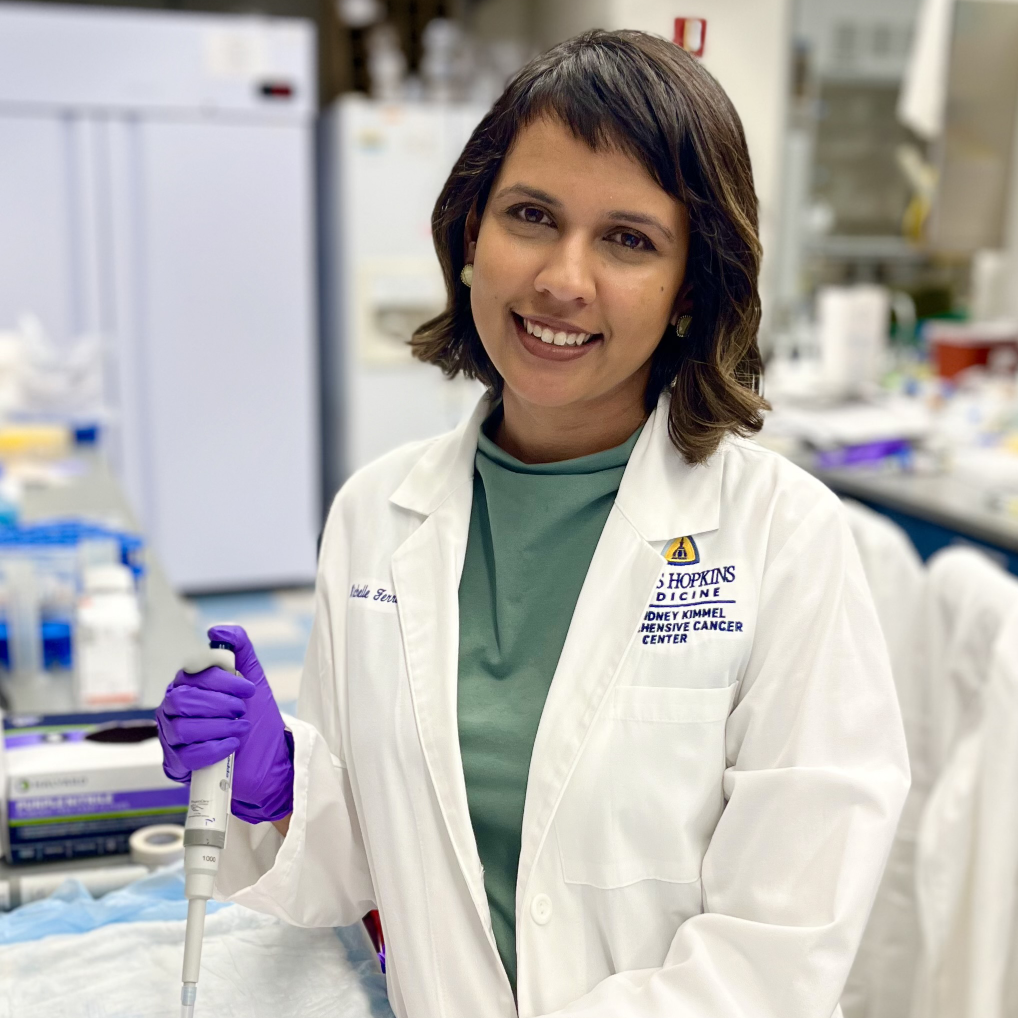
Dr. Ferreira seeks to explore dietary, microbial, and metabolic strategies to treat immune-related colitis (ir-colitis), one of the most common autoimmune toxicities resulting from immunotherapy treatment. Ir-colitis results when the patient’s immune system becomes overstimulated after immunotherapy and attacks the colon, leading to diarrhea and other gastrointestinal symptoms that can result in discontinuation of potentially life-saving immunotherapy. Although ir-colitis can be treated with steroids and other immunosuppressive agents, it remains unclear whether this immunosuppression may affect treatment. Targeting the gut microbiome through dietary intervention is a promising strategy to treat ir-colitis without interfering with cancer treatment. She plans to use a mouse model of ir-colitis to study whether intermittent fasting can be used to treat ir-colitis while simultaneously improving tumor control. She will investigate the specific metabolic and gut microbial changes that are responsible for fasting’s effects on ir-colitis, with the goal of expanding non-immunosuppressive and accessible treatment options for this common immunotherapy toxicity.
Sahana Kuthyar, PhD
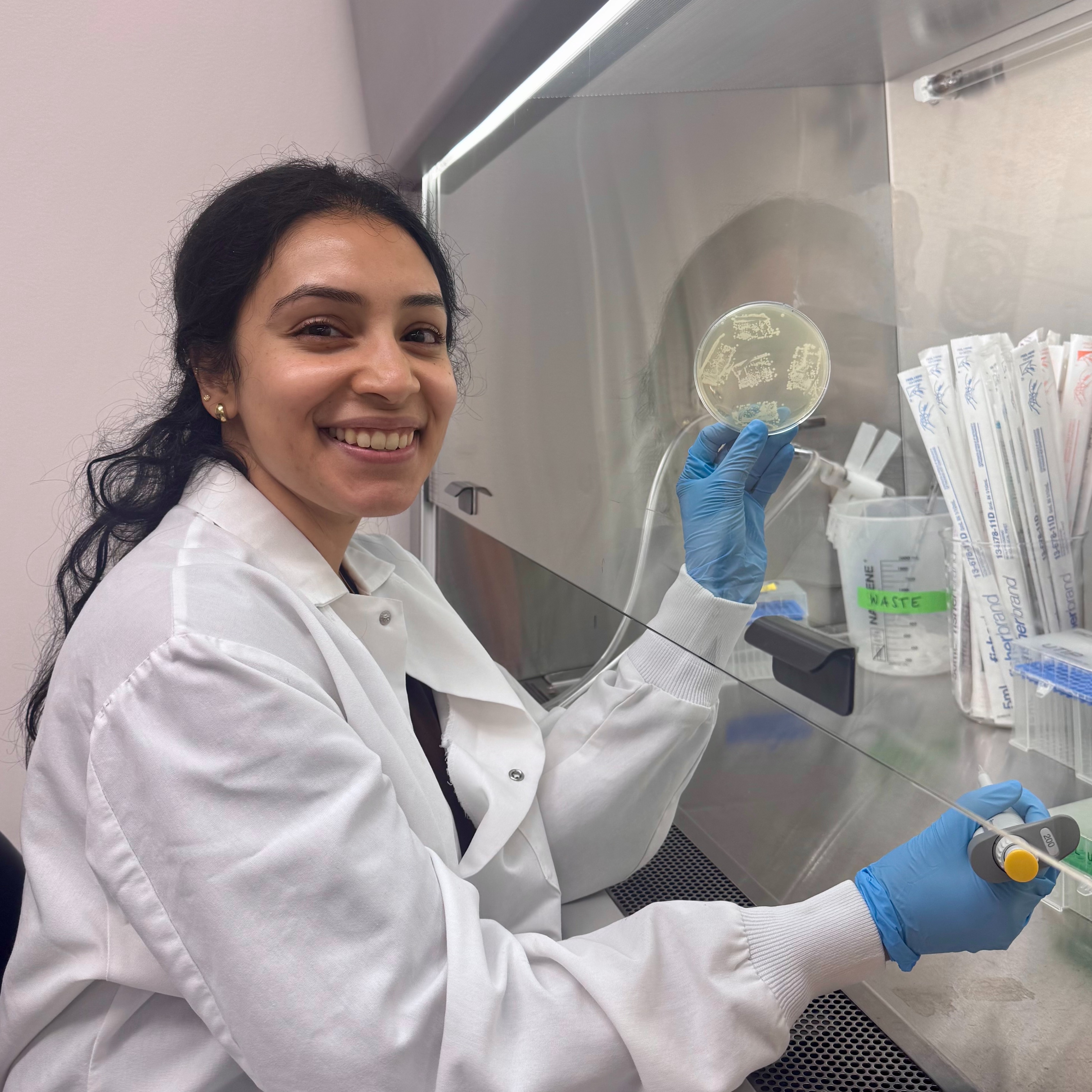
Dr. Kuthyar studies why cancer patients, especially those receiving treatments like chemotherapy or radiation, are at high risk of developing serious lung infections such as pneumonia. While these treatments are essential for killing cancer cells, they also weaken a key part of the immune system that normally helps the body detect and eliminate bacteria. This weakened defense makes patients more vulnerable to infection. At the same time, many hospitalized patients receive supplemental oxygen, which can change the lung environment in ways that help certain bacteria grow stronger and become more aggressive. In cancer patients, these two factors are closely connected: the weakened immune system cannot effectively control bacteria, while the high-oxygen environment actively promotes bacterial survival and virulence. Together, this creates a perfect storm that increases both the risk of contracting pneumonia and severity of disease. This work is relevant to cancers commonly treated with immune-suppressing therapies, including leukemia, lymphoma, and solid tumors such as lung, breast, and colorectal cancer, and aims to identify better ways to predict, prevent, and treat these life-threatening infections.
This project proposes a framework to dissect pneumonia risk in immunocompromised patients using human and mouse models. Dr. Kuthyar will use hierarchical networks to link gene expression and metabolites. Multi-omics factor analysis will capture microbial and immune variation and models trained on human data will be tested in mice, enabling iterative prediction and validation. This approach integrates species harmonization, metabolite prioritization, and network mapping to reveal hyperoxia-driven microbial adaptation and myeloid immune deficits driving pneumonia risk.
Olumide Adesioye
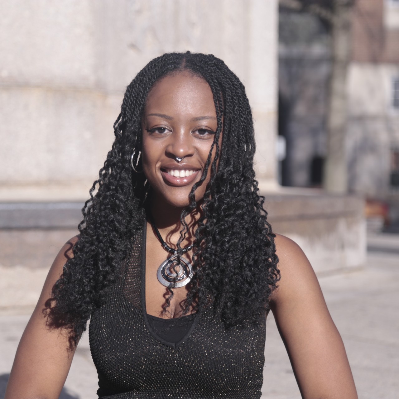
Olumide is a recent graduate of Brown University, where she majored in Immunobiology. Olumide was born and raised outside Chicago by parents who immigrated from Nigeria. After acquiring a passion for biology in her A.P. Bio class in high school, she continued her studies in the biology department at Brown, where she sought out research positions throughout her undergraduate career. Her recent senior honors thesis examined the effects of menopause on immune cell spatial distribution within the endocervix. She is looking forward to learning more about how the immune system can be exploited to fight cancer as a Damon Runyon SPARK Scholar. Olumide is a recipient of the NSF Research Experiences for Undergraduates and a Brown Undergraduate Teaching and Research Award. In her free time, she enjoys weightlifting, roller skating, photography, reading, and hair styling.
Margaret Hu
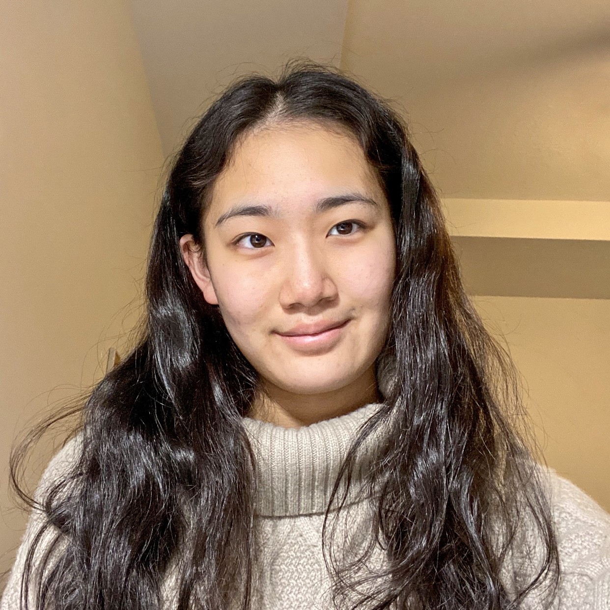
Margaret was born in Wisconsin and grew up in Rochester, Minnesota. She attended Carleton College in Northfield, Minnesota, majoring in Biology and Sociology/Anthropology. Margaret was first exposed to cancer research in Dr. Todd Golub’s lab, where she spent her undergraduate summers studying the tactics employed by leukemia to evade macrophage phagocytosis. This experience fostered her curiosity about cancer’s complex, ever-evolving nature and the wide range of strategies available to target this disease. She aspires to explore human disease at the most fundamental level in order to design elegant and effective new therapies. Margaret’s achievements have been recognized by the Patricia V. Damon Scholar Award from Carleton College. In her free time, she volunteers with her local hospital and patient advocacy organizations and loves to hike, play the piano, and pick up new crafting hobbies.
Aiyina Meng
Aiyina was born in Atlanta, Georgia, to Chinese immigrant parents, and has since lived in four states and three countries. She earned her B.S. in Biology and Biotechnology from Worcester Polytechnic Institute (WPI) in Worcester, Massachusetts, where she developed her research foundation as a trainee in the federally funded Undergraduate Research Training Initiative for Student Enhancement at WPI program. Driven by firsthand experiences watching loved ones navigate serious health challenges, Aiyina has long been motivated to pursue research with meaningful applications in human health and disease. Her research as an undergraduate deepened her commitment to uncovering the molecular mechanisms underlying disease. Aiyina is passionate about translating fundamental science into real-world solutions. Her goal in research is ultimately to contribute to the development of more efficient, targeted cancer therapies. Outside the lab, she enjoys reading, taking long walks, and cooking.
Deahzana Randall
Deahzana was born and raised in Detroit, Michigan. She attended the University of Michigan as a first-generation college student, earning her B.S. in Molecular, Cellular, and Developmental Biology. Her interest in a research career arose from a personal genetic discovery, which sparked her desire to understand the scientific basis of disease. As an undergraduate, Deahzana was selected as a 2025 NK & Irene Cheung Scholar and completed a summer internship in the laboratory of Dr. Santosha Vardhana at Memorial Sloan Kettering Cancer Center, where she explored mechanisms of T cell exhaustion. In the future, Deahzana hopes to attend graduate school with a goal of running her own laboratory and contributing meaningfully to the understanding of tumor progression and immune interactions. Outside of the lab, she enjoys reading, hiking, and watching anime.
Matthew Leventhal, PhD
The X and Y chromosomes play a crucial role in human sex determination. Females have two copies of the X chromosome, while males have one X chromosome and one Y chromosome. In females, the second copy of the X chromosome is silenced early in development, meaning that only one of the X chromosomes is expressed. As a result, mutations on the activated X chromosome are more likely to change cellular functions and in context of cancer, could lead to more rapid disease progression. Dr. Leventhal proposes a novel computational approach to distinguish between the actively expressed and silenced X chromosomes in females. He hopes to use this method to analyze a dataset of over 8,000 tumors to identify potential new drivers and molecular vulnerabilities within 31 types of cancer. He will model whether these alterations can occur in pre-cancerous cells, indicating that they could be targets for early therapeutic intervention.
Dr. Leventhal will develop a computational tool that models the error rate of statistical phasing in bulk whole-genome sequencing and corrects these errors to determine accurate haplotype-specific copy number of all chromosomes. This model integrates genomics with RNA-seq data to determine the active and inactive X chromosome. The subsequent error correction will allow him to perform the first pan-cancer analysis in over 8546 tumors to identify recurrent copy number alterations affecting the active or inactive X chromosome.
Ziyang Zhang, PhD
Dr. Zhang is developing a new form of cancer immunotherapy with improved safety and controllability. Redirecting the immune system to launch attacks on tumor cells has emerged as an extremely promising approach to fight cancer. One such strategy, named bispecific T cell engager antibody (BiTE) has shown remarkable efficacy against blood cancers, but it is also associated with severe toxicity. Using tools of synthetic organic chemistry, he aims to build a “chemical switch” that can be used to rapidly tune the activity of BiTE, thus allowing the circumvention of toxic side effects without diminishing therapeutic potential. The ultimate goal of this project is to develop a cancer immunotherapy that can be safely employed at doses effective for the treatment of solid tumors.
Evan W. Weber, PhD
CAR T cell therapy, in which a patient’s own immune cells are reprogrammed to recognize and kill cancer, has revolutionized the treatment of blood cancers. Unfortunately, however, a significant portion of treated patients relapse, and CAR T cell therapy for aggressive solid tumors has been largely ineffective. A major roadblock preventing this therapy from curing more patients is the gradual loss of CAR T cells’ ability to kill tumor cells, which results in tumor progression or relapse. Dr. Weber aims to develop optimized killer CAR T cells that stay in the fight against cancer. His lab has developed a novel, high-throughput method of analysis that can be used to identify T cell characteristics, genes, and other biological features that enable CAR T cells to serially kill cancer cells. These insights will provide a roadmap for reprogramming T cells with enhanced tumor killing function, paving the way for more efficacious CAR T cell therapies and potentially other cancer immunotherapies for patients in need.
Sarah W. Cai, PhD
Cancer-associated pain can arise directly from tumor growth or as a side effect of chemotherapy drugs. First-line cancer treatments, such as cisplatin and paclitaxel, contribute to pain hypersensitivity by increasing the activity and expression of TRP ion channels—the receptors that detect painful stimuli in sensory neurons and trigger pain sensation. Research thus far has focused on how these receptors look and behave at a single-molecule level (one copy) or at a cellular/organismal level (many thousands of copies per neuron). However, TRP channels are also proposed to operate in nanoscale clusters (tens of copies) that amplify signaling within a sensory neuron. Dr. Cai’s research will use state-of-the-art microscopy techniques alongside biochemical and cell-based approaches to study how receptors are organized on the surface of sensory neurons. She aims to understand how inflammation and injury, including toxicity from chemotherapy drugs, contribute to acute and chronic pain. This work will provide fundamental insights into pain signaling that can inform the development of new pain management strategies. Dr. Cai received her PhD from The Rockefeller University, New York, and her BS from California Institute of Technology, Pasadena.
CONNECT
1.877.7CANCER
info@damonrunyon.org
One Exchange Plaza
55 Broadway, Suite 302
New York, NY 10006![]()
![]()
![]()
![]()
![]()