Bridget P. Keenan, MD, PhD
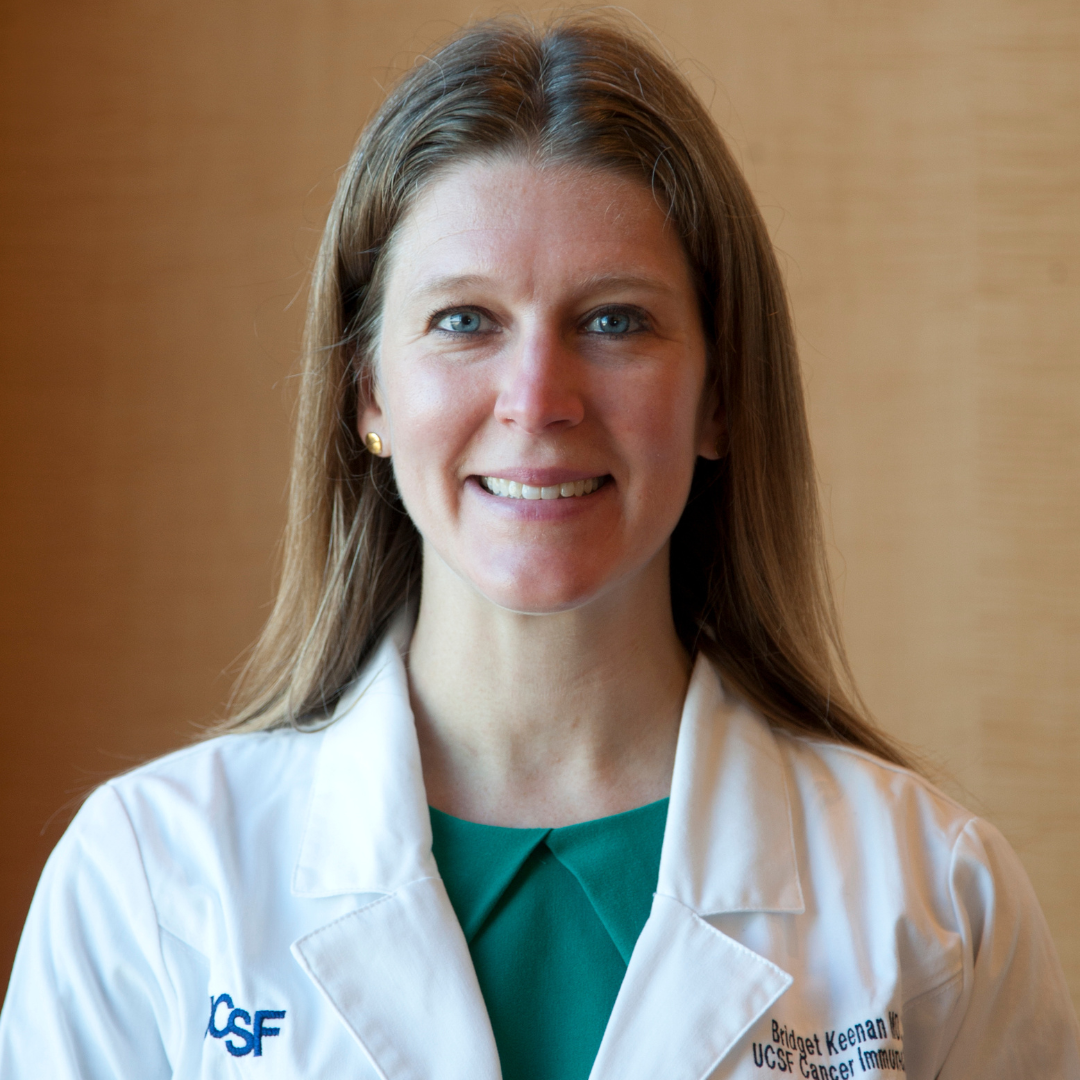
Hepatocellular carcinoma (HCC), a type of liver cancer often caused by liver disease related to viral infections or metabolic disease, is a leading cause of cancer deaths globally. Treating HCC with immunotherapy and targeted therapies shows promise, but liver damage can make these treatments challenging to administer and less effective. Dr. Keenan’s preliminary data suggest that certain immune cells, known as myeloid cells, become suppressive in patients with HCC and worsen liver function. However, it is possible that the correct combinations of immunotherapy treatments could partially reverse this myeloid cell suppression and result in better outcomes for patients with HCC. Dr. Keenan will focus on understanding exactly how liver disease affects the immune system and finding ways to counteract the suppressive effects of myeloid cells. By studying blood samples and liver tissues from patients with HCC undergoing immunotherapy treatment, she aims to identify the best combinations to enhance the immune system’s ability to fight liver cancer. This research could lead to new, more effective treatments for patients with liver cancer, potentially improving survival rates and quality of life.
Tanaya Shree, MD, PhD
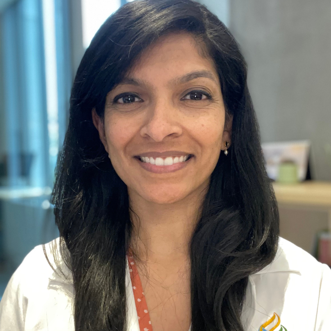
T-cell engaging bispecific antibodies, which bring T cells close to tumor cells and induce them to kill the tumor cell, are a new class of immunotherapy that have demonstrated efficacy in lymphoma and myeloma and are now in development for many other cancers. In diffuse large B cell lymphoma, bispecific antibodies have proven very effective, but approximately 60% of patients derive no long-term benefit. Dr. Shree is working to understand the requirements for generating an effective bispecific antibody response in patients. This knowledge could result in novel improved treatment approaches for patients with lymphoma and inform the design of bispecific T cell-engaging strategies for other types of tumors.
Peter G. Miller, MD, PhD

The goal of Dr. Miller’s research is to determine how mutations in blood cells give rise to pre-malignant blood conditions such as clonal hematopoiesis (CH), which drive the development of blood cancers. To this end, Dr. Miller will study patients with rare inherited diseases and use experimental models in the laboratory. He ultimately seeks to use the data generated through this research to develop new strategies to predict, prevent, and treat highly lethal blood cancers.
Tamar Kavlashvili, PhD

Mitochondria harbor independent genetic material known as mitochondrial DNA (mtDNA). This compact, circular molecule encodes proteins essential for the assembly of the mitochondrial electron transport chain to generate energy in form of ATP. Like nuclear DNA, mtDNA is susceptible to damage and mutations. One of the most common disease-causing aberrations of mtDNA is termed “common deletion.” This aberration disrupts mitochondrial function, resulting in neuromuscular diseases and potentially certain cancers, including colorectal cancer. Due to a lack of tools to modify the mitochondrial genome, researchers currently do not understand the mechanisms behind common deletion. Dr. Kavlashvili [Timmerman Traverse Fellow] aims to investigate by using cutting-edge molecular biology tools to edit and visualize mtDNA genomes. She will then be poised to unravel impacts of this deletion on various tissues, in order to ultimately mitigate its pathological impact. Dr. Kavlashvili received her PhD from Vanderbilt University, Nashville and her BS from University of Iowa, Iowa City.
Rodrigo Gier, PhD

Drug therapies that selectively target proteins that drive the growth of tumor cells are rapidly becoming the standard of care for many cancers. However, tumors are often able to evade inhibition by targeted anti-cancer drugs by activating other proteins, leading to drug resistance. Dr. Gier [HHMI Fellow] is developing a new therapeutic approach that repurposes existing drugs to release highly toxic cargoes, known as payloads, that aggregate in drug-resistant cancer cells and kill them. As a general platform, it is applicable to a wide range of solid and liquid cancers. Dr. Gier received his PhD from University of Pennsylvania, Philadelphia and his BA from Swarthmore College, Swarthmore.
Longyue Lily Cao, MD, PhD

Hepatocellular carcinoma (HCC) is the most common liver cancer and has one of the highest cancer-related mortality rates. Conventional cancer immunotherapies, which largely focus on enhancing T cell activity, are unfortunately effective in only a small minority of HCC patients. Though dendritic cells (DCs) are essential for T cell activation, their potential as an immunotherapeutic target remains poorly understood. Dr. Cao is investigating how a unique, hyperactivated state of DCs can be harnessed to enhance anti-tumor immunity in a genetically engineered mouse model of HCC. Her work aims to uncover how hyperactivated DC responses generate stronger and longer-lasting protection against HCC and hopefully other cancers that are poorly responsive to conventional therapies. Dr. Cao received her MD, PhD from Albert Einstein College of Medicine, Bronx and her BS from Cornell University, Ithaca.
Shaohua Zhang, PhD
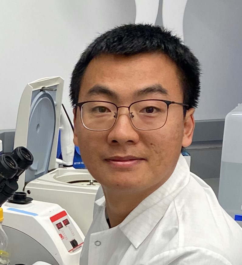
Dr. Zhang [Timmerman Traverse Fellow] aims to engineer T cells with synthetic cell adhesion molecules (synCAMs) to augment current approaches for immunotherapy. This project represents a fundamentally new strategy for CAR T cell engineering that could overcome tumor escape from immunotherapy across multiple forms of cancer. Understanding how synCAMs contribute to CAR T cell efficacy will provide insights beyond cytotoxic CAR T cell therapy; this work could lead to the application of synCAMs in other engineered immune cell therapies under investigation, such as CAR macrophages, CAR natural killer cells, and CAR T regulatory cells. Overall, this approach could lead to CAR T cells that are much more robust to tumor evasion and target antigen expression, and thus much more effective therapeutically. Dr. Zhang received his PhD from University of Chinese Academy of Sciences, Shanghai and his BS from Wuhan University, Wuhan.
Erin M. Parry, MD, PhD

Nicholas P. Lesner, PhD
Ammonia, a waste product of cellular activity, is cleared from the body by the liver and kidneys through a process known as the urea cycle. During the urea cycle, ammonia is converted to urea, and arginine (an amino acid) is generated. When liver cells become cancerous, the urea cycle pathway stops functioning and cancer cells must import arginine from outside the cell. When cancer cells are prevented from importing arginine (via removal of arginine from the diet or genetic removal of the transporter), tumors do not grow, suggesting that arginine is critical for cells. However, the function of arginine in the cell is unclear. Using mass spectrometry and mathematical modeling, Dr. Lesner will identify the fate of arginine as it is metabolized by liver cancer cells in mouse models, and investigate how this is altered by various genetic mutations. Additionally, he will examine how restricting arginine from the diet genetically alters the liver and tumor cells. By understanding how disruption of this metabolic pathway influences liver cancer growth in the context of specific cancer drivers, Dr. Lesner aims to inform new therapeutic strategies. Dr. Lesner received his PhD from The University of Texas Southwestern Medical Center, Dallas and his BA from the University of Wooster, Wooster, Ohio.
Marie R. Siwicki, PhD
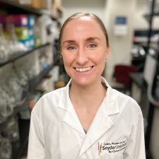
Neutrophils are important anti-microbial cells within the innate immune system. Recently, it has been shown that neutrophils can perform diverse functions, taking on both pro-inflammatory and pro-healing roles in response to tissue injury or insult. Dr. Siwicki's [Dale F. and Betty Ann Frey Fellow] goal is to understand how different neutrophil subtypes or states function to balance inflammatory versus regenerative processes, ultimately influencing tissue health and cancer. This work has the potential to uncover the basis of neutrophils' pro-tumor versus anti-tumor functions and could open the door to therapeutic targeting of specific neutrophil behaviors in order to improve clinical outcomes in cancer. Dr. Siwicki received her PhD from Harvard Medical School, Boston and ScB from Brown University, Providence.
