Yuxuan Chen, PhD
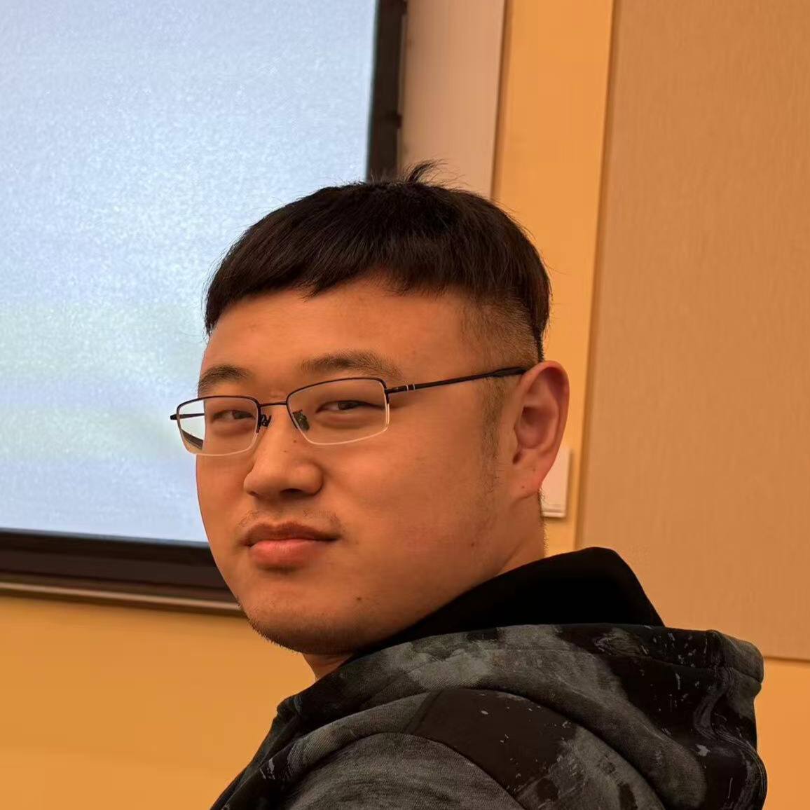
Dr. Chen is genetically re-engineering cancer-infecting viruses so that, once inside a tumor cell, they flip on a built-in “self-destruct” circuit called pyroptosis. This explosive form of cell death not only wipes out the infected cell but also broadcasts an alarm that rallies the immune system against the whole tumor. By pairing this viral upgrade with an ultrasound trigger, Dr. Chen aims to turn treatment-resistant pancreatic cancer—and, ultimately, other solid tumors—into diseases the immune system can eradicate. Dr. Chen received his PhD and MS from Zhejiang University, Hangzhou, and his BS from Southwest Jiaotong University, Chengdu.
Carissa Chan, PhD
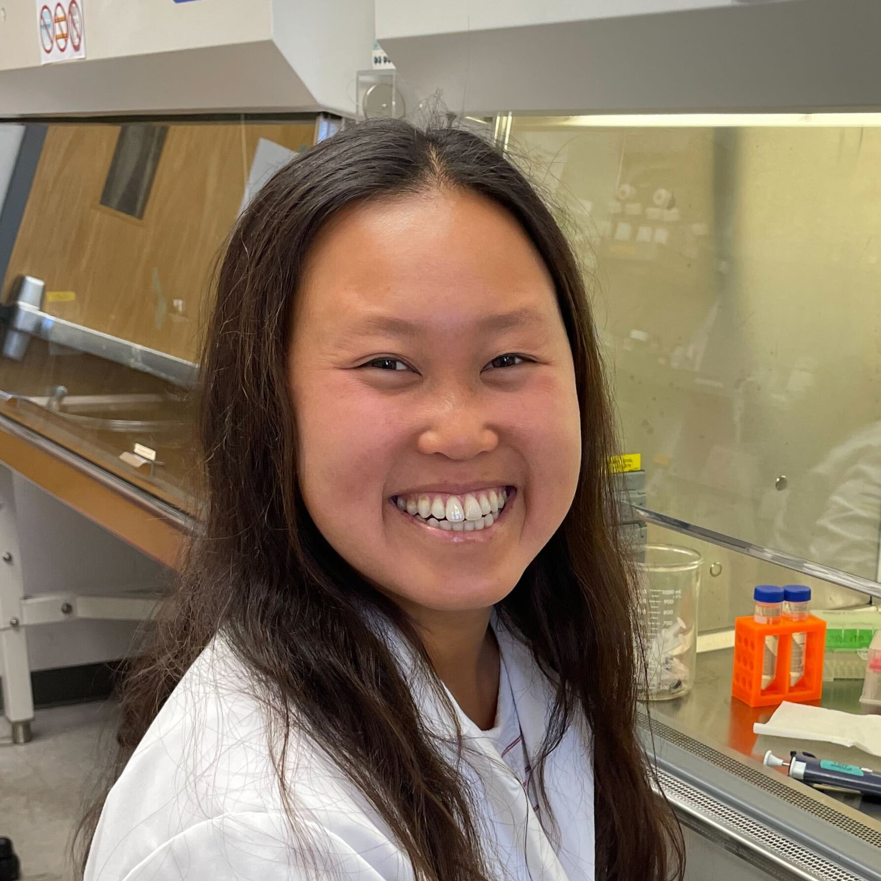
Dr. Chan’s [Sijbrandij Foundation Fellow] research focuses on gamma delta T cells, an unusual and understudied population of immune cells. While gamma delta T cells have strong antitumor activity, they are most highly stimulated not by cancer cells but by signals produced by microorganisms. Dr. Chan’s work examines the mechanisms by which gamma delta T cells detect and respond to leukemia versus pathogenic microorganisms, and how infection with these microorganisms subsequently impacts the trajectory of leukemia. This is particularly relevant to patients undergoing conventional cancer treatments (e.g., chemotherapy) that suppress the immune system, rendering them susceptible to infection. Furthermore, gamma delta T cells are capable of both rapid and long-term responses against their targets, which positions them as a tool to treat initial cancer as well as prevent disease recurrence. Dr. Chan received her PhD from Yale University, New Haven, and her BS from the University of California, Los Angeles.
Paul C. Klauser, PhD
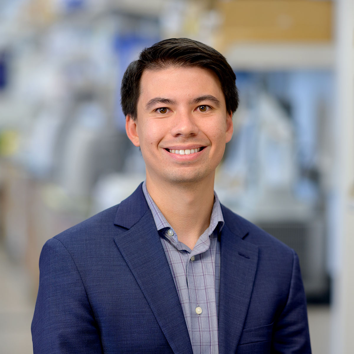
Radiopharmaceuticals, or drugs that contain radioactive forms of chemical elements, have transformed cancer diagnosis and treatment. Radioactive copper and manganese, for example, play a crucial role in PET imaging, while radioactive lutetium is used to deliver targeted radiation to cancer cells. While these radiometals have tremendous potential, however, their application is hindered by a lack of efficient “chelators,” or molecules that can securely bind radiometals in the human body. Computational protein design offers a solution by engineering protein-based chelators optimized for radiometal coordination, stability, and biocompatibility. Using advanced protein modeling, Dr. Klauser [Marilyn and Scott Urdang Quantitative Biology Fellow] will develop chelators for radiometals, improving diagnostic imaging and advancing lutetium-based radiotherapies. While this work is applied to HER2-positive gastric cancer, these strategies have broad applications across various cancer types, ultimately enhancing precision oncology and expanding radiopharmaceutical utility.
This research develops a computational strategy to design stable, compact metal-binding proteins for radiopharmaceuticals, enabling fusion with therapeutic antibodies. Using diffusion models, such as RFdiffusion, thousands of protein backbones are generated for metals like copper and manganese. Sequences are assigned via ProteinMPNN, filtered for stability and binding with AlphaFold 3. For rare lanthanides, symmetric duplication of known binding motifs is used. This approach streamlines the discovery of stable scaffolds for radiopharmaceutical applications.
Ruoyu Wang, PhD
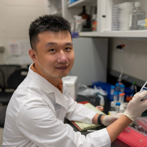
Many cancer mutations occur in regions of the human genome that do not code for proteins. These non-coding regions serve as vital regulators of gene expression; mutations in these regions contribute to various hallmarks of cancer. Elucidating these regulatory elements and their malignant variants is critical for advancing our understanding of cancer biology and fostering precision medicine. Deep learning sequence models can substantially enhance our grasp of the regulatory genome in both health and disease. To this end, Dr. Wang aims to combine generative AI models with single-molecule regulatory genomics to uncover the principles that underlie the cancer regulatory genome at unprecedented resolution and precision.
With single-molecule regulatory genomics, Dr. Wang will develop a deep generative AI model to learn the probability landscape of the single-molecule regulatory genome. By taking any DNA sequence as input, the deep generative AI model can generate diverse configurations of single-molecule chromatin states.
Sohyeon Park, PhD
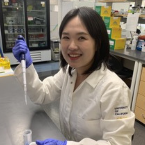
Macrophages are a major component of the body’s first line of defense, acting as sentinel cells that detect and respond to threats. One powerful trait of macrophages is their ability to modulate their response based on previous exposure to stimuli. In cancer, this adaptability can steer macrophages either to fight tumors or to protect them, depending on prior experiences. This phenomenon is often referred to as “macrophage memory.” Though macrophage memory is increasingly recognized as a factor influencing cancer progression and treatment outcomes, the mechanisms that allow macrophages to retain this memory remain unclear. Dr. Park hypothesizes that exposure of macrophages to certain stimuli leads to lasting changes in the structure of their DNA. She will combine both experimental and computational approaches to elucidate how this memory forms and how it affects the expression of immune-related genes. By uncovering the principles of macrophage memory formation, she will lay the groundwork for strategies to reprogram macrophages, potentially enhancing anti-tumor immunity and improving cancer therapy.
Dr. Park will use machine learning–aided genome modeling, leveraging bulk Hi-C data, to reconstruct 3D chromosome structures and validate them with deep learning–based image analysis. This framework will infer single-cell 3D chromosome structure in macrophages. Dr. Park will also quantify nuclear speckle–mRNA spatial relationships using microscopy and develop mathematical models of gene regulation that incorporate transcription factor activity. This integrative approach will reveal how chromosome structure shapes macrophage immune memory and functional response in cancer.
Aaron Zweig, PhD
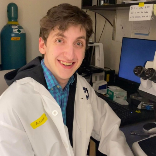
Dr. Zweig [Sijbrandij Foundation Quantitative Biology Fellow] is modeling how gene regulation changes across time and space using advanced geometry, deep learning models, and CRISPR gene-editing technology. With these tools, it is possible to learn how RNA expression evolves in a cell over time and, ideally, predict evolution in regions susceptible to cancerous mutation(s). This work has the potential to apply to many types of cancer but is especially applicable for acute myeloid leukemia (AML). This is in part because there are known “precursor” states—when cells are at higher risk of becoming cancerous—where understanding gene regulation is most impactful, and in part because a common element of AML treatment is a stem cell transplant, which may attack host cells inconsistently in different regions of the same organ.
The project models temporal gene dynamics via stochastic differential equations, parameterizing the drift vector field with linear networks or shallow neural networks to guarantee provable identifiability, trained via differentiation through the adjoint method. Spatial interactions between spot clusters are characterized with graph neural networks, symmetric functions parameterizing summary statistics, and self-attention modules on latent gene embeddings. The latent embeddings are also defined through a variational autoencoder integrating RNA with other modalities.
Akanksha Thawani, PhD

Dr. Thawani studies how so-called “selfish DNA” elements copy and paste themselves within the human genome. Using advanced methods such as cryo-electron microscopy to reveal the atomic structures of various molecules associated with these selfish elements, she aims to delineate their mechanism of mobility. She is also interested in understanding how selfish DNA elements are recognized and silenced within the human genome. Dr. Thawani plans to harness these discoveries to engineer new genome editing technologies to precisely insert large genes at user-specified sites in a variety of human cell types. This general technology will translate directly into new gene therapy tools that will enable treatment of loss-of-function genetic diseases, including many cancer types, and provide a path to improving CAR-T therapies for blood cancers.
Yiyin Erin Chen, MD, PhD

Dr. Chen’s research aims to harness a common skin-colonizing bacterium, present on all our skin, to train the immune system to attack cancer without causing infection or inflammation. This process is known to occur—notably, across an intact skin barrier—but its mechanism is not well understood. Dr. Chen is investigating which skin cells sense these bacteria and transmit the signal to immune cells, and why the immune cells that respond are so effective at killing cancer. Ultimately, she intends to develop a new type of cancer vaccine using engineered skin bacteria to activate immune cells to effectively target and destroy tumors. While the project’s current focus is melanoma, the goal is to apply this therapeutic approach across cancer types.
Xianfeng Zeng, PhD
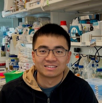
Emerging evidence underscores the profound impact of the gut microbiome, a collection of microorganisms within our digestive system, on cancer. These microorganisms collectively generate various metabolites that can significantly influence cancer progression and treatment outcomes. Dr. Zeng [Fraternal Order of Eagles Fellow] is employing synthetic communities and mouse cancer models to delve into the intricate connections between cancer and the microbiome. His synthetic communities, comprised of over 100 strains, allow for precise manipulation of the microbiome to elucidate the role of specific microbial metabolites in cancer. Additionally, Dr. Zeng is studying community-scale metabolism and using genetically edited strains to design synthetic communities with desired metabolic profiles. These approaches will gain valuable insights into microbiome-cancer interactions and establish a broadly applicable strategy to harness the therapeutic potential of gut microbiome. Dr. Zeng received his PhD from Princeton University, Princeton and his BS from Tsinghua University, Beijing.
Teng Gao, PhD

Hematopoietic stem cells, which are found in the bone marrow and give rise to all other blood cells, maintain lifelong blood production and immune function. Due to their remarkable ability to regenerate the entire blood system, medical uses of HSCs have provided cures for many previously incurable diseases, including blood cancers. However, several unanswered questions limit our ability to full harness their therapeutic potential for cancer treatment. What regulates HSC regeneration? Why does their function decline with age? How does HSC behavior vary in healthy individuals? Using cutting-edge single-cell analyses and computational biology, Dr. Gao [HHMI Fellow] aims to identify the molecular and cellular factors involved in HSC regeneration, as well as possible targets for enhancing their regenerative potential. This work could enable significant improvements in stem cell-based therapies for cancer treatment. Dr. Gao received his PhD from Harvard University, Cambridge and his BS from Washington University, St. Louis.
